Original Source: https://kendricklabsinc.wordpress.com/2022/11/07/2d-electrophoresis-all-about-its-methodology-and-working-principle/
Electrophoresis is a process that applies electric current to molecules in a buffer to separate the DNA matrix. It allows for analysis of the nucleic acid structure. Because electrophoresis is used in laboratory research, it can also be used as an experimental DNA and RNA analysis technique. In 2-D electrophoresis, you can apply different conditions that will allow for the separation of molecules according to their size or charge distribution on the proteins within them. For example, electromigration uses low applied voltage with high frequency, while electroosmosis employs high applied voltage and low frequency of application.
How Does It Work?
Depending on the mode of application and conditions, the electrophoresis can separate the DNA molecules from each other. In 2-D electrophoresis, you can apply different conditions that will allow for the separation of molecules according to their size or charge distribution on the proteins within them. Two critical factors that influence electrophoresis are pH and current strength. The glycerol (water) buffer is one of the most common buffers in 2-D electrophoresis. The glycerol neutralizes the acidic or basic pH of the sample, making it an excellent buffering solution.
Working Principle:
Electrophoresis is a technique for separating charged particles, and you can hire a reliable service at Kendrick Laboratories. It uses electrical resistance to separate molecules. In a gel, DNA fragments get separated by their size. The rate of migration depends on the ionic strength and pH of the buffer, as well as the strength of the electric field and the shape and dimensions of the gel matrix. Samples are applied to the wells in a gel matrix and subjected to an electric field. The DNA fragments move through the pores of the gel, with the most negative charges moving first.
Methodology:
The three main steps of 2-D electrophoresis involve SDS-PAGE (sodium dodecyl sulfate-polyacrylamide gel electrophoresis), Ethidium bromide staining, and autoradiography. Electrophoresis is performed by mixing equal amounts of agarose or acrylamide solution with 10x TBE buffer, then pouring it between two glass slides and placing them in a tank filled with TBE buffer. DNA is then placed in the wells, and 10x TBE buffer is poured over it. A current is applied to electrophoresis. DNA migrates through the pores of the gel matrix uniformly. DNA fragments are removed from the wells and soaked in a sodium dodecyl sulfonate (SDS) solution an SDS-protein complex replaces them. Negative-charged RNA and protein can be removed, whereas DNA remains intact due to its positive charge. The gel is then incubated with a primary antibody against DNA.
Sample Solubilization:
DNA is diluted to a low concentration and is not stable in the sample solution. It is mixed with SDS and forms a solution that migrates through the gel matrix and will be transferred to another agarose or acrylamide solution. After reducing the sample concentration, it is loaded onto a gel. DNA loading involves placing gel pieces into small wells and pouring excess buffer. After sample loading, it passes through the holes in the gel matrix and into the next chamber until no more plasma can pass through any gap between well plates.
Isoelectric Focusing:
To perform IEF, samples are diluted in an SDS-water system and loaded onto a gel. DNA is bound to a column and washed with a buffer solution. Sodium dodecyl sulfate (SDS) causes the separation of protein and DNA negatively charges them by adding negative charges to the ends of the molecules that cause them to counteract each other. In this way, DNA can be concentrated on one side of the gel while protein remains in the same place on the opposite side. After washing, it is stained with ethidium bromide.
Exposure of Gel to X-ray Film:
After washing, proteins are stained with ethidium bromide. This process is also known as chromatography, and the staining binds to DNA. Images of gels can be obtained through autoradiography or laser scanning and recorded using a digital or photographic device. DNA fragments move into the wells due to the electric field and are separated based on size or shape.
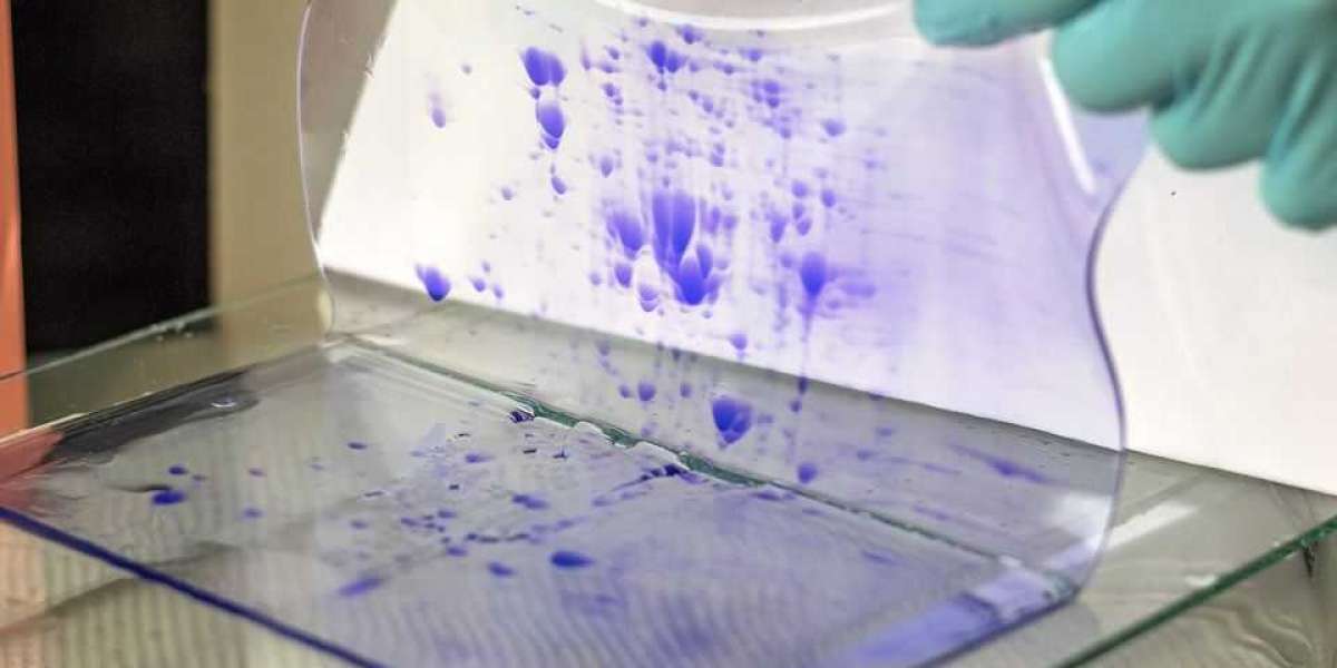
Representative Results:
The results of 2-D electrophoresis show how well gel electrophoresis works by separating the mixtures into various bands for analysis. Each band on the gel represents a specific molecule that had been separated by size during gel mobility. They also showed how efficient 2-D electrophoresis is by allowing scientists to separate molecules by size with the added advantage of determining the molecule type.
Applications:
Two-dimensional gel electrophoresis (2DGE) is an efficient and powerful technique for analyzing complex protein mixtures. Conventional two-dimensional electrophoretic methods are mainly used in oligonucleotide mapping, genomic DNA cloning, and human genome project. In research, chromatographic separations are used as the first step in experiments like proteomics, where separation can be more beneficial than subsequent identification because it enables the investigation of large biomolecules with simple detection procedures.
Conclusion:
Various applications of gel electrophoresis are present in academic and industrial areas. For example, it is used in research to type bacteria, find their origin and investigate, characterize and identify many different species of micro organism. Other uses include identifying food-borne microorganisms, characterization of mycobacteria, and pathogen identification from human or animal samples.